Wrist Arthroscopy & Wrist Pain
Role of Wrist Arthroscopy for Wrist Pain
Patients who complain of wrist pain usually describe pain which interferes with their daily activities, work or sports. Some may report a history of specific wrist injury, the commonest mechanism of injury being fall on outstretched hand. They will usually be prescribed a trial of conservative treatment which includes non-steroidal anti-inflammatory medications, steroid injections, rest and splinting initially for an average period of 3-6 months. Investigations performed would include X-rays and magnetic resonance imaging (MRI).
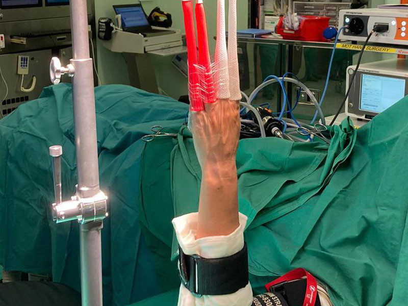
When initial management fails, we usually proceed to perform wrist arthroscopy under general or regional anesthesia. Wrist arthroscopy has progressed from a newly described technique since the 1970s to become an essential diagnostic and therapeutic tool in the armamentarium of every surgeon treating various wrist disorders. It is useful in defining the patterns, combinations and extent of soft tissue injuries which are often more extensive than clinically suspected. In treating patients with chronic wrist pain recalcitrant to conservative treatment with wrist arthroscopy, we found many patients having concomitant intercarpal ligament (scapholunate & lunotriquetral ligament) injuries as well as triangular fibrocartilage complex (TFCC) injuries. Other findings included synovitis and chondral lesions such as lunate chondromalacia. It is not uncommon to find double or triple pathologies.
Many procedures can be performed arthroscopically such as synovectomy, radiofrequency ablation, debridement of any chondral defects, repair of peripheral TFCC tears and even limited fusion. Postoperatively the wrist was placed in a brace or splint for 6 weeks and started on occupational therapy for range of motion and grip-strengthening exercises.
Wrist arthroscopy has a valuable role in the treatment of chronic wrist pain. It allows direct visualization of the wrist joint and therefore identification of multiple partial injuries in the wrist, such as ligament attenuations and chondromalacia, not readily apparent on other imaging modalities. Many surgeons regard it as the gold standard investigation in the diagnosis of carpal instability, especially as it can detect isolated tears of intrinsic ligaments and demonstrate degenerative joint changes. Wrist arthroscopy also enables one to treat the pathology during the intervention. Its disadvantages are the surgical risks, the need for regional or general anesthesia as well as its costs.
The main limitation of arthroscopy is that not all abnormalities identified by arthroscopy are necessarily pathologic or responsible for the patient’s symptoms. Evaluation of the results of treatment is also difficult because treatment of coexistent injuries may be responsible for the alteration of symptoms. However, many studies have proven wrist arthroscopy to be a safe, reliable with a favorable impact upon the outcomes, especially in relieving wrist pain.
 11 May 2024
 08 Apr 2023
 21 Jun 2023